Distal Femur Fracture Overview & Treatment Options in Chicago
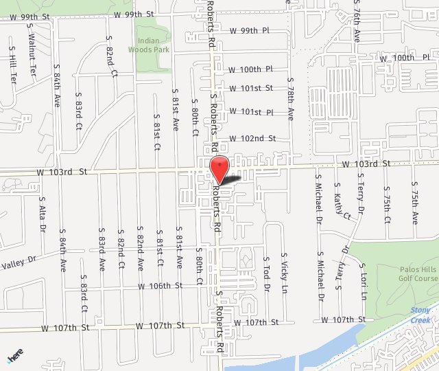
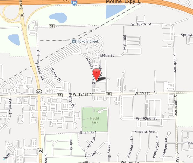
With convenient locations in Palos Hills and Mokena, IL
A distal femur fracture is located just above the knee joint. It takes a significant amount of force to break this bone and if it occurs, you need to seek immediate medical attention. These fractures can vary in severity based on a number of factors. Dr. Bedikian is a highly skilled practitioner with over 20 years of experience treating serious orthopedic injuries, including distal femur fractures.
Types of Distal Femur Fractures
Transverse fracture: This type of fracture is when the fracture extends straight across the bone.
Comminuted fracture: If the bone breaks in a way that results in many different fragments, it is known as a comminuted fracture.
Intra-articular fracture: This is a complex injury that extends into the joint and also involves damage to the cartilage.

Causes of Distal Femur Fractures
Injuries like falls from significant heights or motor vehicle accidents are common causes of distal femur fractures.
In older individuals, poor bone quality due to aging can make the bones more susceptible to fracture during low-impact events, such as falling from a low height.
Symptoms and Diagnosis
Common symptoms include:
- Severe pain when putting weight on the knee
- Swelling
- Bruising
- Knee pain that extends to the thigh
- A knee that appears deformed, or crooked
Diagnosing a distal femur fracture begins with a physical exam, where Dr. Bedikian will examine the injured area to determine if it is an open or closed fracture. Because this type of injury is often associated with other injuries to different parts of the body, your overall condition may also be assessed during your exam.
X-rays are commonly used to gather additional information about the fracture. They can also reveal the precise location as well as the type of fracture within the femur. An additional benefit of X-rays is that they can identify any associated injuries in nearby bones and joints.
You may also receive a CT scan, which generates cross-sectional images of the injured site. This provides valuable insights regarding the severity of the fracture. This imaging technique can also show the number of bone fragments, which helps Dr. Bedikian determine the most appropriate treatment for you.
Modern Practice with Modern Solutions
Our state-of-the-art clinic is home to advanced tools and techniques such as robotic-assisted surgery and arthroscopy for joint procedures.
Treatment Options
Most distal femur fractures require surgery. The healing process for a broken femur typically takes about two months, with some cases requiring more than six months to fully heal. Surgery may not take place right away, especially if there is significant swelling or the patient is not fit for surgery. In these cases, Dr. Bedikian may utilize an external fixator to align the bones and reduce pain until the swelling subsides enough to safely perform surgery. However, in cases where the bone fragments have broken the skin, surgery is usually done immediately.
Distal femur surgery involves incisions on the front, outside, and inside of the knee and thigh, potentially extending along the entire side of the thigh in certain cases. Metal plates and screws are then used to realign the bones.
Seeking Medical Care
If you suspect a distal femur fracture, it is important to seek immediate medical attention. Contact our office today to schedule an appointment with Dr. Bedikian.


